A mammogram is a specialized X-ray exam used to examine breast tissue for signs of cancer. Doctors generally recommend these screenings for women starting at age 40, and regular exams help detect cancer early when it is most treatable. Because medical science is always evolving, recent technological advancements have made these screenings more accurate than ever before. While traditional exams were effective, new tools allow radiologists to see tissue more clearly.
Exploring AI-supported Mammography
Artificial intelligence, or AI, can be used to support radiologists during the review of a mammogram. When it is used to read a mammogram result, this technology acts as a second reader in addition to the radiologist. The computer analyzes the images, but a human doctor makes the final diagnosis. The software uses deep learning algorithms that have studied thousands of images, and it identifies potential abnormalities that the human eye might miss.
AI technology also improves efficiency for the medical team. When radiologists use AI to assist them in reading images, they can work faster without increasing false positives. Reading hundreds of images per patient takes time, but AI cuts that reading time significantly.
Understanding Tomosynthesis Imaging
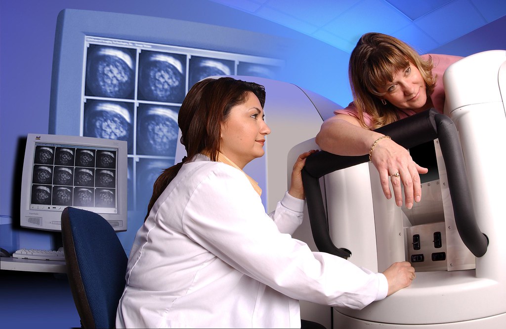
Digital breast tomosynthesis, or DBT, creates a three-dimensional picture of the breast. The X-ray tube moves in an arc around the breast, and it captures images from multiple angles. When the radiologist has multiple angles to look at, they can see more detail than with a traditional 2D mammogram.
The procedure feels very similar to a standard exam for the patient. A technologist positions the breast on a platform and compresses it with a plastic paddle. While the breast remains compressed, the machine moves in an arc to take the X-rays. The computer then digitizes these pictures into a clear 3D representation. The process takes only a few seconds, but the resulting images provide a wealth of information.
This technology is particularly beneficial for women with dense breast tissue. Dense tissue appears white on an X-ray, and cancer also appears white. Digital breast tomosynthesis allows the radiologist to see more detail in dense tissue. DBT more effectively distinguishes between overlapping tissue and actual tumors. Studies show that 3D mammography increases cancer detection rates in dense tissue, and it reduces the chance of false positives.
Recognizing Screening Benefits
Regular screenings serve as an early defense against breast cancer fatalities. Early detection is the main goal of screenings. You might not feel a lump or notice abnormalities, but the X-ray can find them. When cancer is found early, treatment is generally less invasive. Doctors can treat the disease more effectively before it spreads, and this proactive approach offers the best chance for a cure.
There are two main types of breast exams that work together. A clinical breast exam involves a doctor checking for physical lumps; a mammogram looks for changes inside the tissue that are too small to feel. Using both methods provides a comprehensive view of your health. You can also perform self-exams at home to monitor any changes you notice in your own body.
Get a Mammogram Today
Technology in mammograms has advanced, and screenings are now more precise than they were in the past. Because these tools improve screening results, they make mammograms even more effective at finding cancer in the early stages. To learn more about mammograms and their benefits, consult a women’s health provider near you.